INTRODUCTION


Hepatitis B virus (HBV), hepatitis C virus (HCV) and human immunodeficiency virus - HIV (HIV) are anthroponotic viruses - affect cells only of the human body and, therefore, cause disease only in humans. Experimental models of these infections do not exist. There are also no cultured cell cultures on which invitro could adequately study the cytopathogenic properties and viability of these viruses.

Characteristics of hepatitis B, hepatitis C and HIV viruses.

Hepatitis B virus (HBV) is a DNA-containing virus, its replication occurs mainly in human hepatocytes. The hepatitis B virus also has lymphotropic properties — the ability to infect and replicate in human lymphocytes.
Hepatitis C virus (HCV) is the RNA-containing virus with the property of infecting and replicating in both hepatocytes and human lymphocytes.
Human immunodeficiency virus (HIV) refers to enveloped retroviruses and is the RNA-containing virus. The main target cells for damage and replication are CD4 lymphocytes.
Consequently, the common property of HBV, HCV and HIV is their lymphotropism - the ability to penetrate and replicate in lymphoid cells.

II PREREQUISITES FOR THE CREATION OF A METHOD FOR DETECTING AND EVALUATING THE VIABILITY OF LYMPHOTROPIC VIRUSES HBV, HCV AND HIV.

Studies on the issues of the clinic, pathogenesis, treatment and diagnosis of chronic viral hepatitis, conducted by employees of the Laboratory for the study of the chronic infectious process (Head of Prof. Gulyamov N.G.) in the USRIEMID of the Ministry of Health of the Republic of Uzbekistan, as well as the analysis of numerous scientific literature data, revealed that in chronic HBV and HCV infections, the severity of secondary immunodeficiency has no natural correlation with the nature and severity of inflammatory and destructive processes in liver tissue. Along with this, there are isolated reports of the possibility of persistence of HBV and HCV, as well as HIV, in mononuclear blood cells, in particular in lymphocytes and macrophages. However, no researcher has paid attention to the assessment of this fact from the standpoint of the pathogenesis and clinic of viral hepatitis.

It was found that with HBV and HCV infections, inflammatory processes in the liver develop simultaneously with all the signs of hepatitis that follow from this, as well as a secondary immunodeficiency state, expressed in varying degrees of T-lymphopenia and B-lymphopenia, in disproportions of regulatory subpopulations of T-lymphocytes (T-helpers and T-suppressors), in a decrease in the immunoregulatory index (IRI), as well as in the phenomena of dysgammaglobulinemia. The degree and severity of immunodeficiency did not have a natural relationship with the severity of the pathological process in the liver. In patients with chronic HBV and HCV infections, after time, the intensity of the progression of the pathological process in the liver tissue may be different - from mild to severe, but, regardless of this, there is a stable and steady aggravation of secondary immunodeficiency.

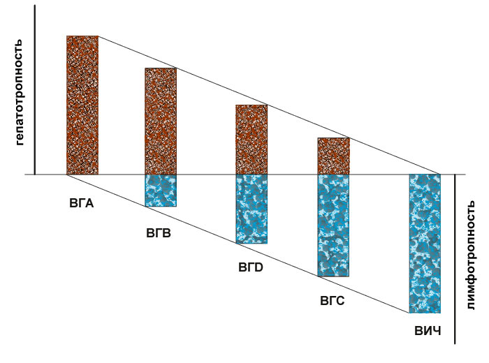
Dissociation of the degree of liver tissue damage and the depth of secondary immunodeficiency in various nosological forms of chronic viral hepatitis gave reason to assume that hepatitis and secondary immunodeficiency in HBV and HCV infections are combined processes, mutually aggravating, but are not mutually conditioning: that is, HBV and HCV, along with hepatotropy, have a pronounced lymphotropic property – an indirect property of forming secondary immunodeficiency. At the same time, differences in the clinical manifestations of liver tissue damage and the depth of immunodeficiency in HBV and HCV infections are caused by differences in the severity of hepatotropic and lymphotropic properties of these viruses. It is the differences in the degree of severity of the hepatotropic and lymphotropic properties of viruses that cause differences in pathogenesis, clinical manifestations and the nature of the effect of antiviral therapy in chronic HBV and HCV infections at different time periods of the disease.

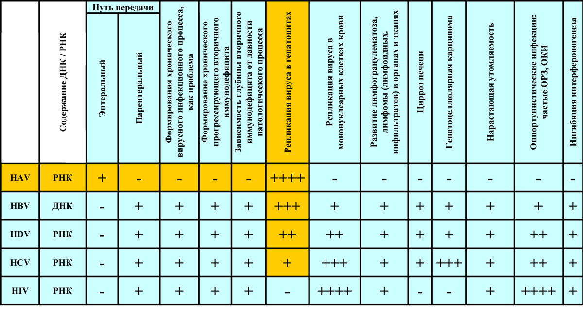
The analysis of classical ideas about the clinic and the outcomes of various diseases of viral etiology in humans revealed a pattern: a cyclic course with complete elimination of the pathogen and the outcome of recovery is naturally characteristic of diseases caused by viruses, including the hepatitis A virus (HAV), which do not have a specific tropism relative to the cells of the immune system - lymphotropy; the formation of a chronic, recurrent and constantly progressing infectious process is naturally a characteristic feature of human diseases caused by viruses with a specific tropism relative to immune cells, that is, lymphotropicity, of which HIV infection is a striking example.

Drawing the analogy between clinical and immunological indicators, variants of the clinical course, manifestations of the effect of antiviral therapy and disease outcomes in HAV, HBV and HCV infections, we found that HAV has mainly only hepatotropic properties and only in isolated cases gives chronization, and even then only when viral hepatitis develops against the background of already existing chronic liver pathology or secondary immunodeficiency. However, HAV does not show obvious lymphotropic properties. The absence of secondary immunodeficiency due to the direct hepatotropic action of HAV allows the immune system to implement an adequate antiviral protective reaction, resulting in the elimination of HAV from the body and complete recovery. HBV and HCV, having a hepatotropic effect to varying degrees, simultaneously have a direct lymphotropic, i.e. secondary immunodeficiency forming effect.

It is the defeat of immune cells and the development of secondary immunodeficiency in HBV and HCV infections that limits the ability of the immune system to implement a full-fledged antiviral reaction of the macroorganism, resulting in the formation of a chronic infectious process that resembles that of HIV infection. The identity of the lymphotropic properties of HBV and HCV, as well as HIV, in addition to the formation of secondary immunodeficiency, is also confirmed by the commonality of their epidemiological features, transmission mechanisms, the development of concomitant opportunistic infections (frequent acute respiratory infections, acute respiratory infections) and, especially, the development of lymphogranulomatosis in various tissues of the body. The development of lymphogranulomatosis in various organs and tissues of the body is considered to be inherent in viral lesions of the lymphoid cell system (Table 1, Fig. 1).

Considering the lymphotropic properties of HBV, it is not difficult to assume that HBV antigens (HBsAg), regardless of their titer in serum, can persist permanently and significantly in high concentrations in the cytoplasm of lymphoid elements. It was this phenomenon that we used to increase the reliability and exclude false negative results in the ELISA analysis.

Using ELISA analysis, we conducted studies on simultaneous testing for the presence of HBsAg in the blood serum and lymphocyte contents of patients with suspected viral hepatitis. It was found that out of the total number of examined patients in 17% of cases, on average, the result of the ELISA analysis of blood serum was false negative, since high HBsAg titers were detected in the lymphocyte contents of the same individuals. These results irrefutably prove the lymphotropic properties of HBV and are certainly of great practical importance for increasing the reliability and reducing false negative results when testing blood for the presence of HBsAg using ELISA analysis.

So, the lymphotropic properties of HBV and HCV, as well as HIV, that is, their ability to penetrate and replicate in human lymphocytes, are the established fact.

Based on the above, to develop the method for detecting and evaluating the viability of HBV, HCV and HIV, we used lymphotropic properties — the ability of these viruses to penetrate and persist intracellularly in healthy human lymphocytes during their joint incubation in vitro.

III THE PRINCIPLE OF THE METHOD FOR DETECTING AND ASSESSING THE VIABILITY OF LYMPHOTROPIC VIRUSES HBV, HCV AND HIV


The method of detecting and assessing the viability of viruses is based on the use of lymphotropic properties – the ability of HBV, HCV or HIV to penetrate and persist in the lymphocytes of a healthy person.

During incubation of biological material with a suspension of lymphocytes of the healthy person, in the presence of HBV, HCV or HIV in the biological material, viruses penetrate into the cytoplasm of lymphocytes. Further, lymphocytes are concentrated in a small volume by centrifugation and destroyed by freezing.

The presented method consists of the following stages: obtaining a suspension of lymphocytes from the blood of a healthy person, incubation of lymphocytes with the biological material under study (plasma, blood serum, etc.) precipitation of lymphocytes by centrifugation, washing of lymphocytes in saline solution, precipitation of lymphocytes and dissolution in a small volume (500.0 ml of saline solution), destruction of lymphocytes by freezing, thawing. The contents of the destroyed lymphocytes are subjected to ELISA or PCR examination. Detection of viruses in the contents of lymphocytes by ELISA or PCR indicates the presence and viability of viruses in the biological material under study.

IV METHOD OF DETECTION OF LYMPHOTROPIC VIRUSES HBV, HCV AND HIV IN LYMPHOCYTES OF PATIENTS AND BLOOD DONORS.


The method makes it possible to detect viruses in patients and blood donors even at low virus titers in plasma or serum.

  • 1. In the examined person, 5-6 ml of vein blood is taken from the ulnar vein into a test tube containing 2 ml of saline solution and 2-3 drops of heparin.
  • 2. Lymphocytes are isolated from whole heparinized blood by separation in a ficoll-verografin solution with a density gradient of d = 1.077 g/ml according to the method of F.Y. Garib et al. (1995). In the process of centrifugation, all shaped blood elements, except lymphocytes, penetrate through the thickness of the ficoll-verografin gradient and are located under it. The blood plasma remains above the gradient. In the thickness of the gradient, a kind of cloudy ring ("cloud") is formed, consisting of a pure suspension of lymphocytes. This ring with lymphocytes is carefully sucked off with a pipette and transferred to a clean centrifuge tube.
  • 3. Next, a double washing of lymphocytes in 10.0 ml of saline solution is carried out, followed by.
  • 4. After the last centrifugation, the filler liquid is removed.
  • 5. The sediment containing lymphocytes is diluted in 500 ml of saline solution, transferred to Eppendorf and placed in the freezer of a household refrigerator for 17-18 hours. With slow freezing, the lymphocyte membrane is destroyed and the contents of the cytoplasm are released into solution.
  • 6. After 17-18 hours, the Eppendorf is removed from the freezer, thawed at room temperature.
  • 7. The contents of the Eppendorf are subjected to PCR or ELISA examination for the presence of HBV, HCV or HIV markers.


This method allows to concentrate viruses from 5-6 ml of plasma or serum in 500 ml of volume, increase the titer of viral particles above the threshold of sensitivity of the ELISA or PCR method. This helps to exclude false negative results of ELISA or PCR when testing blood donors or patients for their infection with HBV, HCV or HIV.

V METHOD FOR DETECTING LYMPHOTROPIC VIRUSES IN BIOLOGICAL MEDIA WITH A VERY LOW VIRUS TITER CONTENT.


  • Obtaining suspension of lymphocytes of healthy person.
  • Healthy human volunteers are tested for infection with HBV, HCV and HIV by the ELISA method. In tests, lymphocytes of healthy people are used as target cells only with negative test results.
  • To obtain a sufficient amount of lymphocyte suspension, blood is taken from a healthy person (volunteer) in the morning on an empty stomach from the ulnar vein in an amount of 30-40 ml. Next, 7-8 ml of blood is transferred to centrifuge tubes containing 2 ml of saline solution with 3 drops of heparin. The mixture in the test tube is thoroughly mixed;
  • The isolation of lymphocytes from whole heparinized blood is carried out by separation in a solution of ficoll-verografin with a density gradient of d = 1.077 g / ml according to the method of F.Y. Garib et al. (1995). To do this, 2 ml of ficoll-verografin solution is poured into clean centrifuge tubes, heparinized blood is carefully layered on its surface and the tubes are centrifuged at 1500 rpm for 20 minutes. During centrifugation, all shaped blood elements, except lymphocytes, penetrate through the thickness of the ficoll-verografin gradient and are located under it. The blood plasma remains above the gradient. In the thickness of the gradient, a kind of cloudy ring ("cloud") is formed, consisting of a pure suspension of lymphocytes. This ring with lymphocytes is carefully sucked off with a pipette and transferred to a clean centrifuge tube;
  • Next, a 2x-3x washing of lymphocytes in 10.0 ml of saline solution is carried out, followed by centrifugation.
  • After the last centrifugation, the filler liquid is removed. Sediments containing lymphocytes are diluted in 500 ml of saline solution and mixed. A suspension of lymphocytes from all tubes merge into one tube. The suspension of lymphocytes can be stored for no more than 1 day at a temperature of + 4oC.Incubation of lymphocytes with the biological medium under study.
6-7 ml of the studied biological medium (plasma, donor blood serum or others) is poured into 2-3 clean test tubes, which is subjected to ELISA or PCR testing for infection with HBV, HCV or HIV.

  • 500-600 ml of a healthy person's lymphocyte suspension is added to each tube, the contents of the tubes are thoroughly mixed and placed in a thermostat at 37°C for 6-8 hours. Every 0.5-1 hour, the contents of the test tubes are shaken and mixed. During incubation, in the presence of viruses in the biological environment under study, the latter, due to their lymphotropic properties, adhere to the membrane surface and penetrate into the cytoplasm of lymphocytes.
  • After incubation, the test tubes are removed from the thermostat, 2 ml of saline solution is added to them, the mixture is mixed and subjected to centrifugation. During centrifugation, lymphocytes are deposited at the bottom of the tubes.
  • After centrifugation, the settling liquid is removed from all the tubes, the precipitates are transferred to one tube. Next, lymphocytes are washed twice in saline solution.
  • After the last centrifugation, the settling fluid is removed from the test tube, the sediment containing lymphocytes is diluted with 600 ml of saline solution and transferred to the epindorph. Epindorf is placed in the freezer of the refrigerator. With slow freezing, the lymphocyte membrane is destroyed and the contents of the cytoplasm are released into solution.
  • After 17-18 hours, the epindorf is removed from the freezer, thawed at room temperature.
  • The contents of the epindorph are subjected to PCR or ELISA examination for the presence of HBV, HCV or HIV markers.
This method makes it possible to concentrate viruses from 18-20 ml of biological medium in 500 ml of volume, to increase the titer of viral particles above the threshold of sensitivity of the ELISA or PCR method. This helps to exclude false negative results of ELISA or PCR when testing biological media (plasma, serum of donated blood and others) for infection with HBV, HCV or HIV.

VI METHOD FOR ASSESSING IN VITRO VIABILITY OF LYMPHOTROPIC VIRUSES HBV, HCV AND HIV.

  • Obtaining suspension of HBV, HCV or HIV. To obtain suspension of viruses in patients infected with HBV, HCV or HIV monoinfections, with a previously known high viral load, blood is taken from the ulnar vein. Virus-containing serum is isolated from whole blood, which is subjected to quantitative PCR examination to confirm the presence of HBV, HCV or HIV and to determine the titer of viruses. The virus-containing serum is stored frozen in a minus refrigerator at a temperature below - 25 ° C.
  • Obtaining suspension of lymphocytes of a healthy person.
  • Healthy human volunteers are tested for infection with HBV, HCV and HIV by the ELISA method. The tests use lymphocytes of healthy people only with negative test results;
  • To obtain sufficient amount of lymphocyte suspension, blood is taken from a healthy person (volunteer) in the morning on an empty stomach from the ulnar vein in an amount of 30-40 ml. Next, 7-8 ml of blood is transferred to centrifuge tubes containing 2 ml of saline solution with 3 drops of heparin. The mixture in the test tube is thoroughly mixed;
  • The isolation of lymphocytes from whole heparinized blood is carried out by separation in the ficoll-verografin gradient with a density of d = 1.077 g / ml according to the method of F.Y. Garib et al. (1995). To do this, 2 ml of ficoll-verografin gradient is poured into clean centrifuge tubes, heparinized blood is carefully layered on its surface and the tubes are centrifuged at 1500 rpm. During centrifugation, all shaped blood elements, except lymphocytes, penetrate through the thickness of the ficoll-verografin gradient and are located under it. The blood plasma remains above the gradient. In the thickness of the gradient, a kind of cloudy ring ("cloud") is formed, consisting of a suspension of lymphocytes. This ring with lymphocytes is carefully sucked off with a pipette and transferred to a clean centrifuge tube;
  • Next, a 2x-3x washing of lymphocytes in 10.0 ml of saline solution is carried out. After the last centrifugation, the filler liquid is removed. Sediments containing lymphocytes are diluted in 500 ml of saline solution and mixed. A suspension of lymphocytes from all tubes merge into one tube. The suspension of lymphocytes can be stored for no more than 1 day at a temperature of + 4oC.


In vitro evaluation of the ability of HBV, HCV or HIV viruses to penetrate the cytoplasm of human lymphocytes.

  • HBV, HCV or HIV containing plasma is removed from the minus refrigerator, thawed at room temperature.
  • The equal volume (300 ml each) of virus-containing plasma and lymphocyte suspension is transferred to a clean test tube using an automatic pipette, the contents are mixed and put for incubation (incubation of viruses with invitro lymphocytes) in a thermostat at a temperature of +37°C for 6-8 hours. The contents of the test tube are mixed by shaking every 1.5-2 hours. During incubation, only viable viruses that exhibit cytopathogenic effects penetrate into the cytoplasm of lymphocytes.
  • Washing of lymphocytes from plasma. Next, the test tube is removed from the thermostat. 6-8 ml of saline solution is added to it, mixed and centrifuged at 1500 rpm/min. There is a deposition of lymphocytes to the bottom of the tubes. The filler fluid is removed. Next, a 2x-3x washing in saline solution and precipitation of lymphocytes is performed.
  • After the last centrifugation and removal of the infusion fluid, the suspension of lymphocytes (sediment) is diluted in 500 mcl of saline solution and transferred to the epindorph.
  • After that, for the destruction of lymphocytes, epindorph is placed in the freezer of a household refrigerator for 17-18 hours. With slow freezing, the destruction of lymphocyte membranes occurs.
  • The supraventricular fluid from the epindorph is sucked out and subjected to quantitative PCR examination for the detection of DNA or RNA viruses in the cytoplasm of lymphocytes that were previously contained in the blood plasma from patients. Evaluation of results.
  • A positive result of a PCR study for the presence of RNA or DNA viruses in the contents of the cytoplasm of lymphocytes indicates the preservation of the viability of viruses, that is, their ability to penetrate and persist in human lymphocytes in vitro.
  • A negative result of PCR examination for the presence of RNA or DNA viruses in the contents of the cytoplasm of lymphocytes indicates the loss of viability (inactivation) of viruses (after exposure to antiviral factors of chemical or physical nature), that is, the loss of their ability to penetrate and persist in human lymphocytes in vitro.


VII THE RANGE OF APPLICATIONS OF THE METHOD FOR DETECTING AND ASSESSING THE VIABILITY OF LYMPHOTROPIC VIRUSES HBV, HCV AND HIV

  • Blood transfusion stations: to increase the reliability of detection of lymphotropic hepatitis B (HBV), hepatitis C (HCV) and human immunodeficiency virus (HIV) in biological media (donor blood, plasma and other components) containing viruses in concentrations below the threshold of sensitivity of ELISA or PCR methods.
  • Diagnostic laboratories: to increase the reliability and exclude false negative results when testing for HBV, HCV or HIV infection in the blood of donors or patients with virus content in concentrations below the threshold of sensitivity of ELISA or PCR methods.
  • Pharmaceutical companies: : to assess the viability of HBV, HCV or HIV viruses after exposure to chemical and physical antiviral factors developed and offered by manufacturers in order to inactivate viruses at various facilities.
  • The method makes it possible to eliminate errors in the diagnosis of patients, when testing donated blood and its components for infection with lymphotropic viruses HBV, HCV or HIV. The method makes it possible to objectively assess the effects of chemical or physical factors on the viability (pathogenicity) of lymphotropic viruses.
  • The method of detecting and evaluating the viability of lymphotropic viruses has no analogues.
  • This method is registered by the Ministry of Health of the Republic of Uzbekistan.

Author: Gulyamov Nariman Gulyamovich, Doctor of Medicine, Professor